Floor View Skull Dorsum Sellae

Specialised Projections Of The Skull Radiology Key
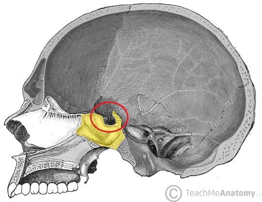
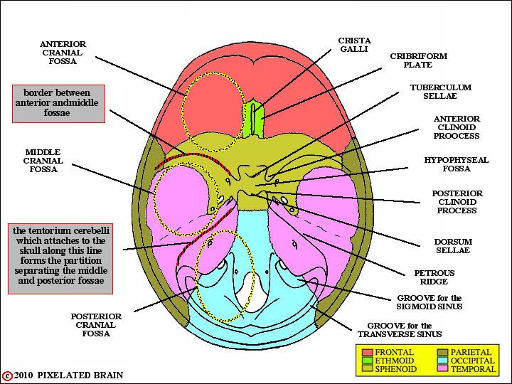
Middle Cranial Fossa Boundaries Contents Teachmeanatomy

Landmarks For Cephalometric Analysis S Sella Center Of Sella Download Scientific Diagram
Anatomy Of The Skull Base And Related Structures Elements Of Surgical Anatomy Neupsy Key

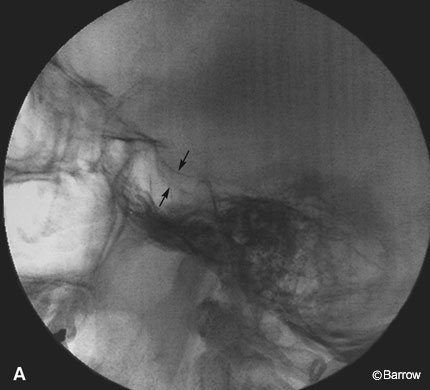
Skull X Ray Lateral View Note Enlargement Of Pituitary Fossa Loss Download Scientific Diagram
Pixelated Brain Module 1 Section 1 The Skull
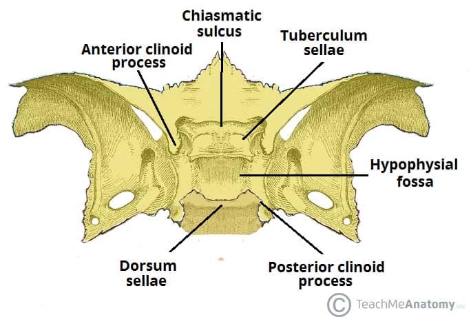
It serves as a cephalometric landmark.
Floor view skull dorsum sellae.

The Axial Skeleton Flashcards Quizlet
Sphenoid Bone Location Structure Function Teachmeanatomy
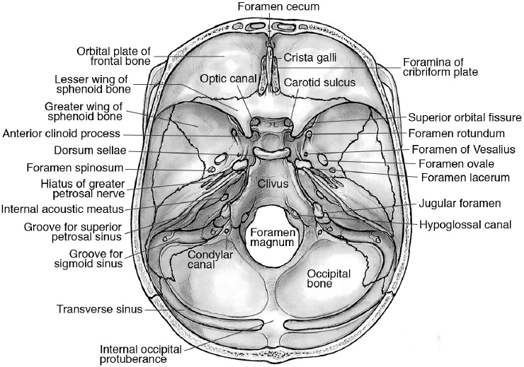
Skull Foramina Fissures And Contents Kenhub

Sphenoid Bone
Source : pinterest.com
